The impact of spatial biology on biomarker research
• The spatial architecture of tissue samples can strongly influence disease pathology, progression, and treatment response. Several recent studies have outlined the importance of studying the spatial context of tumor samples to predict treatment response.
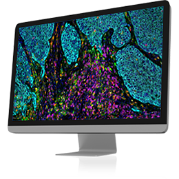
• Traditional immunohistochemistry (IHC) preserves spatial context but is limited to 2 to 3 biomarkers per sample. Next-generation sequencing (NGS) enables the analysis of multiple biomarkers but the spatial context of the tissue is lost. Multiplex imaging (multiplex IHC) addresses these limitations by enabling the analysis of multiple biomarkers in a tissue section while preserving their spatial context.
• The SpITR core implements this high-plex tissue profiling technology to study tumor heterogeneity and microenvironment.
PhenoCycler-Fusion 2.0 Technology
The PhenoCycler-Fusion 2.0 technology uses antibodies conjugated to a proprietary library of oligonucleotides called Barcodes. This enables customizable panels of up to 40+ PhenoCycler-Fusion Assays to be combined for a single tissue staining reaction.
PhenoCycler Services & Information
Services
SpITR offers highly multiplexed imaging using the PhenoCycler platform. The technology allows for detection of a maximum of 40 protein targets in the same tissue section at single cell resolution, with quantitative data for each protein. Since 2018 SpITR has been part of the PhenoCycler early access program. Now, the technology is available as a service as collaborative support with cost recovery for the consumables.
Pricing
• Slide Staining: $750.00 / slide
• Antibodies: $28.00 / antibody*
• Cycles: $12.00 / cycle
Projects totaling $6,000 or more are eligible for 50% OSTR subsidy up to an $8,000 subsidy maximum per PI per fiscal year.
Tissue Types
SpITR has extensive experience with imaging fresh frozen tissues, as well as working knowledge and experience with FFPE and TMAs.
- Mouse Fresh Frozen
- Human Fresh Frozen
- Human FFPE
- TMAs (tumor microarrays)
Tissue Preperation & Sectioning
Users are responsible for harvesting and preparing tissue sections onto coated coverslips for PhenoCycler experiments. Use poly- L-Lysine coated coverslips for fresh frozen and Vectabond or APES treated coverslips for FFPE tissues. The coated coverslips are provided by SpITR.
For best quality sections, special care should be taken both for collection and fixation of tissues. For these steps follow general histology procedures (Akoya’s manual for freezing specimens and manual for frozen tissue sectioning).
The following website has technical notes for best practices for tissue processing:
Tissue size: The recommended tissue thickness for fresh frozen is 8 um (max 10 um) and for FFPE the recommendation is 4-5 um. The tissue should not exceed 1.6 x 1.6 cm. The tissues should be placed in the middle of the coverslip as much as possible and free of folds and wrinkles.
Molecular Histopathology and Histoserv have experience with this type of tissue sectioning.
Number of sections: SpITR requests 3-5 tissues sections for each sample and recommends having H&E staining on adjacent slides. If you need help with slide scanning, SpITR coordinates with LGCP for automated scanning and images are uploaded into HALO image analysis platform for viewing and collaboration.
Detected targets by PhenoCycler: The maximum number of targets that can currently be detected is 40 – driven by the number of commercially available barcodes. The antibody panel comprises of commercial and custom conjugated antibodies.
The commercial antibody panel is different based on the species and tissue type: mouse FF, human FF, human FFPE (SpITR Target List)Tissue staining, imaging, and processing: After finalizing the antibody panel, SpITR staff performs tissue staining and imaging following recommended best practices from Akoya Biosciences, using commercial PhenoCycler microfluidics connected to a Keyence microscope. The whole tissue section or different regions are imaged using a 20X objective acquiring multiple Z-stacks. Imaging time depends on the area of the tissue and number of targets and can take 1-2 days per sample. Raw images are processed to generate aligned and improved 2D stitched 16-bit tiff images.
Results delivered: A summary power point slide deck generated from PhenoCycler processor with experimental details. Stitched tiff images for each individual signal, as well as composite images, are uploaded into HALO for viewing, qualitative and single cell level quantitative analysis. Tile by tile raw or processed images are delivered upon request.
Data Analysis: For basic analysis, SpITR recommends HALO (IndicaLabs) image analysis software for area, single cell level and spatial analysis. The cloud version of the software is available for investigators through CBIIT and includes AI enabled tools for tissue classifications and cellular/nuclear segmentation. SpITR staff provides guidelines and can direct users to training resources for PhenoCycler image analysis. Single cell level quantitative data can be exported as csv files for further analysis in any third-party software.
If interested in PhenoCycler-Fusion 2.0 technology for a project: For questions and to discuss new projects please contact us via our group email: CCRSPITR@mail.nih.gov Next, we will schedule a meeting to discuss your needs, clarifying the sample type, number and antibody panel needed and ask you to submit a project proposal at https://spitr.ccr.cancer.gov/projm/. Upon project approval you will be asked to fill out the sample submission form and prepare the samples.
Advantages of PhenoCycler-Fusion 2.0 Technology
• Provides full spatial context and is not limited to just regions of interest (ROI)
• Provides single-cell resolution down to 600 nm or 250 nm depending on microscope objective used (20X and 40X respectively)
• Single-step staining and gentle fluorophore removal preserves the sample for ROI analysis
• Simple, benchtop fluidics system that is cost-effective and simple to implement in any research lab

